Love Your Heart: A Cleveland Clinic Podcast
Love Your Heart: A Cleveland Clinic Podcast
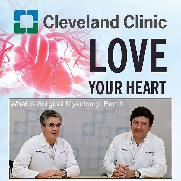
What is Surgical Myectomy: Part 1
A septal myectomy is an open-heart surgical procedure to treat hypertrophic cardiomyopathy, a thickened heart muscle. This allows blood to flow better through the heart. Dr. Nicholas Smedira and Dr. Juan Pablo Umaña provide an overview of what may cause a patient to need a septal myectomy. Part 2 will further discuss treatment, surgery and recovery.
Learn more about the specialized Hypertrophic Cardiomyopathy Center at Cleveland Clinic
https://my.clevelandclinic.org/departments/heart/depts/hypertrophic-cardiomyopathy
Learn more about Hypertrophic Cardiomyopathy
https://my.clevelandclinic.org/services/hypertrophic-cardiomyopathy-treatment#featured-provider-panel
Listen to Part 2: https://my.clevelandclinic.org/podcasts/love-your-heart/what-is-surgical-myectomy-part-2
Announcer:
Welcome to Love Your Heart, brought to you by Cleveland Clinic's Sydell and Arnold Miller Family Heart, Vascular & Thoracic Institute. These podcasts will help you learn more about your heart, thoracic and vascular systems, ways to stay healthy and information about diseases and treatment options. Enjoy!
Nicholas Smedira, MD, MBA:
Hi, I'm Nick Smedira, and I'm here with Dr. Juan Umaña. We're here to talk about septal myectomy, which Juan, is a really dynamic changing practice in cardiac surgery. There's so much going on in the field of hypertrophic cardiomyopathy, so it's a real exciting time for the management of patients so that we have so many things now in our armamentarium. It's really exciting.
Juan Pablo Umaña, MD:
It is. I think that it's one of the most exciting fields right now, wouldn't you say? Not only from a medical treatment perspective with all the new options available, but as you've pointed out several times recently, surgery is still the backbone of treatment for these patients. I think there are a lot of questions as to what is hypertrophic cardiomyopathy and when it should be treated.
Given that you're the expert in the field, not only at the Cleveland Clinic, but worldwide, why don't you tell us a little bit [about] what patients should be operated on, what hypertrophic cardiomyopathy, particularly the obstructive kind, is, and when patients should be thinking of seeing a cardiologist and perhaps even being referred to a surgeon.
Nicholas Smedira, MD, MBA:
There's a lot to unpack there, but for starters, hypertrophic cardiomyopathy is a genetic disorder. Interestingly, we thought the genetic disorder occurred in about 1 in 500, but in fact, it may be as common as 1 in 250. If you do the math, that's a lot of patients that are potentially affected by this gene.
Now, not everybody that has the gene will get that thick part of the heart of the septum, but many patients will. What we need is to understand what's going on with the heart in terms of what we call obstruction. The blood has to get out of the heart, and the pathway is bordered by muscle on one side and the mitral valve on the other. To effectively exit- the blood to get out of the heart, there has to be a space for that. In hypertrophic cardiomyopathy and in some diseases of the mitral valve, that space gets too narrow and the blood can't get out.
To really understand what's going on, a patient with symptoms such as dizziness, maybe they even passed out, shortness of breath especially, interestingly, after a meal. That's one of the hallmark signs of hypertrophic obstructive cardiomyopathy is shortness of breath after a meal, which is very interesting.
The patient then is seen by an imaging specialist who does an echocardiogram. Often, you need to run on a treadmill to make sure the obstruction is identified, and then it's critical with either echo or MRI or combination of those exams to understand how much of the problem is from the muscle, which gets thick, or the mitral valve, which could be in the wrong position.
It can be too long, any assortment of combinations. That's where visiting a center of excellence that has the experience to identify what exactly is going on is critical. Then of course, it's important to have the surgical expertise to do what operation is necessary.
What I do commonly is just shave the muscle. It's a relatively simple operation, and your expertise is the mitral valve and the mitral valve pathology, and what can be going on. Here, we've developed a number of unique operations that can help the mitral valve perform better without having to replace it.
Our goal is to try to do things so the patient has their own valve, shaved muscle, and avoid a mitral valve replacement. That's from the start through surgery that we've developed here, and we also do in Weston down in Florida.
Juan Pablo Umaña, MD:
If we take it from the top, simplify, I think that one of the things that is fascinating about the outflow tract, or what it takes for the blood to get out of the left ventricle, it's not only the ventricle contracting, but the mitral valve interacting with the muscle, as you said.
When that muscle gets very thick, that mitral valve may just get almost sucked into that outflow tract. That's when the expertise of a multidisciplinary team comes in, which is exactly what you've been working on here for a long time with imaging cardiologists, with echocardiographers, with MRI, as well as a clinical cardiologist that's dedicated to hypertrophic cardiomyopathy.
The surgery itself sounds very complicated and very complex, but as you get down to it, Nick, it's really a relatively simple operation if you understand the concept, and you've done it several thousand times, right?
Nicholas Smedira, MD, MBA:
Correct.
Juan Pablo Umaña, MD:
How often would you think that the mitral has to be repaired or intervened in hypertrophic cardiomyopathy?
Nicholas Smedira, MD, MBA:
I think if you look at the big picture of obstruction, blood getting out of the heart, I'd say the vast majority of the time it's the muscle and not the valve. By cutting the muscle thinning that septum, you open up enough pathway to let the blood out in the mitral valve that behaves normally. Now we have a little bit of a referral bias because we've written a lot about the mitral valve being the primary culprit for obstruction.
Cardiologists around the country, around the world send patients to us who have thinner septums. They're not very hypertrophied, but they still have obstruction. When we started down this pathway, we saw this, and as cardiologists began to more intensely investigate patients with symptoms, because in our minds as we just started to discuss, it was all about thickness of the septum, hypertrophy. If you didn't see hypertrophy on the echocardiogram, you didn't think of obstruction because at rest, the obstruction might not be there because it often only happens with exercise or activity or a heavy meal.
Juan Pablo Umaña, MD:
Which is why you were saying that it's important to do an exercise [stress test]-
Nicholas Smedira, MD, MBA:
To provoke.
Juan Pablo Umaña, MD:
Yeah, provoke the obstruction.
Nicholas Smedira, MD, MBA:
There's been so many patients, which is so regrettable when you hear the story, that have said, "I have had symptoms since I've been in high school. I couldn't do gym class. I went into the Marines and I barely made it through bootcamp. I was so short of breath." But then when they examined them at rest, they have no murmur, the echocardiogram looks perfectly normal, but if you had them do 50 jumping jacks in your office, or we often have Valsalva, which can provoke the obstruction, you get the obstruction, but calm things down, you don't see anything.
That was very, very difficult. Patients are told they have asthma, people are told that they have anxiety, they have panic attacks, they see neurologists for dizziness, and it's this obstruction that may only be present during when the heart's contracting. We identified patients that didn't have a lot of hypertrophy and asked just the fundamental research question: if you don't have hypertrophy and you're obstructing, what's causing the obstruction?
Of course it was the mitral valve, and then the question was: why is the mitral valve doing this? Because that then leads to thinking of surgical techniques that can intervene. Putting a stitch may not be the answer once you understand the anatomy and the physiology. We started to look at how long is the leaflet? Because some mitral leaflets can be very excessively long, and that's causing them to flip up.
I think potentially a couple causes. One is that when you're a fetus in utero, when the heart twists, the papillary muscles end up rotating in a position that puts them in the outflow path, so we've developed a technique to move them out of the outflow path.
Juan Pablo Umaña, MD:
Just for anybody who might be watching and listening to this, wondering what papillary muscles are, the mitral valve looks almost like a parachute, right?
Nicholas Smedira, MD, MBA:
It is.
Juan Pablo Umaña, MD:
It's held down into the ventricle by these cords that are attached to two muscles. You're saying that sometimes, those can be slightly rotated and as a consequence, be in the way of the flow of blood.
Nicholas Smedira, MD, MBA:
That's what we've observed. We've focused on them for a bit to pull them back down away from the septum. We have a couple of techniques to do that. I also think as we age, and maybe as we gain some weight, we lift up the heart a little bit as our diaphragm comes up, and that changes the angles of which the blood can get out of the heart. That predisposes patients to some obstruction.
I think that's an explanation for why would somebody in their late 60s or 70 all of a sudden develop obstruction, and then we do genetic testing and they don't have any of the genes that we know cause obstruction. I think what happens is they may have a little high blood pressure, they may have this change that occurs as we age, and the next thing that you know that you're predisposed to obstruction.
These techniques that we've developed, these observations that we made have led to us being more attuned to diagnosing the disorder and then having a couple of techniques to take some muscle, repair the mitral valve to open up the path. It's been 25 years of working together in a center where we not only do the surgery, but we study the details of what we do, we combine the imaging aspect of it, and we've come up with, I think a fairly good way to then answer your question, how often does this occur in our referral where we repair the valve maybe 10% of the time?
Juan Pablo Umaña, MD:
Really, it's a small percentage, and the reason for that is we have a very deep understanding of the whole disease process and the pathophysiology of the obstruction, right?
Nicholas Smedira, MD, MBA:
That's correct.
Announcer:
Thank you for listening. We hope you enjoyed the podcast. We welcome your comments and feedback. Please contact us at heart@ccf.org. Like what you heard? Subscribe wherever you get your podcasts, or listen at clevelandclinic.org/LoveYourHeartPodcast.